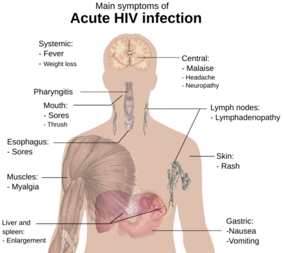
Lymphocytic pleocytosis
Elevated CSF lymphocytes signal nervous system infection or inflammation.
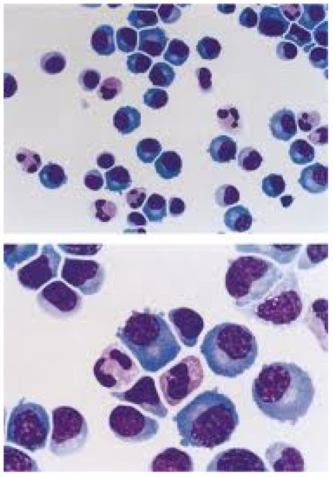
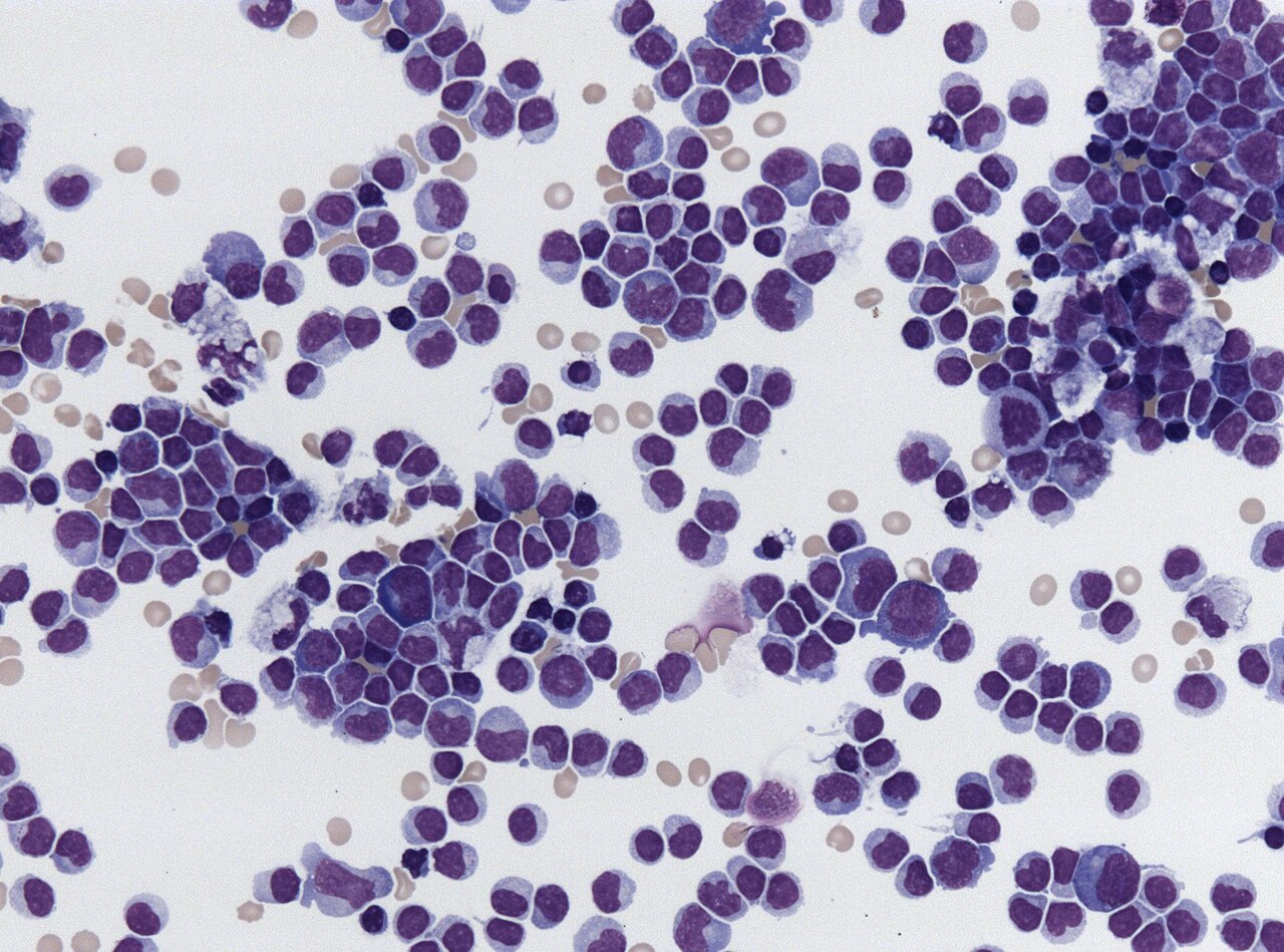
Lymphocytic pleocytosis is an abnormal increase in the number of lymphocytes in the cerebrospinal fluid (CSF). It is considered a sign of infection or inflammation within the nervous system and is encountered in neurological diseases such as pseudomigraine, Susac's syndrome, and encephalitis. While lymphocytes make up roughly a quarter of all white blood cells in the body, they are generally rare in the CSF, with normal levels below 5 white blood cells per μL.
- field
- Neurology, Clinical Pathology
- known_for
- Abnormal increase of lymphocytes in cerebrospinal fluid indicating nervous system infection or inflammation
- normal_CSF_WBC_count
- 0-5 cells per μL
- pleocytic_lymphocyte_range
- 10 to over 1,000 cells per μL
- associated_diseases
- Pseudomigraine, Susac's syndrome, encephalitis, viral meningitis, Mollaret's disease, Lyme disease
Lore & Background
Lymphocytic pleocytosis is detected through a lumbar puncture followed by clinical analysis of cerebrospinal fluid. The presence of elevated lymphocytes is often accompanied by increased cerebrospinal protein concentrations and pleocytosis of other white blood cells. In healthy individuals, only 0-5 white blood cells per μL are normally present, but in pleocytic settings, lymphocyte counts can jump to more than 1,000 cells per μL. Mild cases begin when lymphocyte counts enter the range of 10-100 cells per mm³. Increases to more than 500 cells per mm³ can cause the CSF to appear cloudy.
Reader's Guide
Lymphocytic pleocytosis is a key diagnostic marker for several neurological conditions. Its detection via lumbar puncture helps diagnose diseases such as subarachnoid hemorrhage, multiple sclerosis, and various types of meningitis. The condition is generally the result of an immune response to neurovascular inflammation, often triggered by viral infections. Treatment focuses on the underlying cause: antiviral drugs like aciclovir and valacyclovir for herpes infections, or immunosuppressive drugs like prednisone for autoimmune responses. While a normal WBC count does not rule out disease, the presence of lymphocytic pleocytosis provides critical clinical information for diagnosis and management.
Did You Know?
- Lymphocytes make up roughly a quarter of all white blood cells in the body but are generally rare in the CSF.
- In pseudomigraine patients, CSF lymphocyte concentrations have been shown to range from 10 to 760 cells per mm³, with a mean of 199 ± 174 cells per mm³.
- More than 90% of patients with herpes simplex virus encephalitis are found to have lymphocytic pleocytosis of varying levels.
- In Susac's syndrome, an autoimmune response damages blood vessels of the brain, retina, and cochlea, leading to hearing and vision loss along with elevated CSF lymphocytes and proteins.
The Lymphocyte Threshold in Cerebrospinal Fluid
Lymphocytic pleocytosis describes an abnormal elevation of lymphocytes within the cerebrospinal fluid, a condition that typically signals infection or inflammation somewhere within the nervous system. Although lymphocytes constitute roughly one quarter of all circulating white blood cells throughout the body, they are exceedingly scarce in healthy CSF, where total white blood cell counts normally remain below five cells per microliter. When pleocytosis develops, lymphocyte concentrations can surge dramatically, sometimes exceeding one thousand cells per microliter. Mild presentations are generally recognized when counts fall between ten and one hundred cells per cubic millimeter, while readings above five hundred cells per cubic millimeter may render the fluid visibly cloudy during laboratory examination. These elevations frequently travel alongside raised cerebrospinal protein levels and increases in other white blood cell populations. The underlying driver is most often an inflammatory immune response, particularly one triggered by viral invasion of neural tissue. The condition has been documented across a spectrum of neurological disorders, including encephalitis, Susac's syndrome, and pseudomigraine, making it a critical diagnostic marker in neurology.
Diagnostic Detection and Clinical Interpretation
The primary route to identifying lymphocytic pleocytosis is a lumbar puncture, during which a sample of cerebrospinal fluid is withdrawn for laboratory analysis. Clinicians do not rely on white blood cell counts alone; rather, they integrate the fluid's visual appearance, opening pressure, glucose concentration, and protein levels to build a comprehensive diagnostic picture. This combined approach helps distinguish conditions such as subarachnoid hemorrhage, multiple sclerosis, and the various forms of meningitis. Importantly, a lumbar puncture that returns a white blood cell count within the normal zero-to-five range does not definitively exclude disease, meaning a negative result must be interpreted cautiously alongside the patient's clinical presentation. In pseudomigraine research, for instance, lymphocyte concentrations have been recorded anywhere from ten to seven hundred sixty cells per cubic millimeter, with a mean of roughly one hundred ninety-nine. Imaging studies in these patients revealed normal CT and MRI scans, yet thirty of forty-two individuals showed abnormal EEG findings, including unilateral or bilateral slowing. This underscores that pleocytosis detection and neurological assessment must proceed in tandem.
Disease Associations and Pathological Mechanisms
Lymphocytic pleocytosis appears across a diverse array of neurological conditions, each with distinct pathophysiology. In viral encephalitis, the infection inflames brain tissue and disrupts neurological function, producing fever, confusion, amnesia, personality shifts, paralysis, seizures, and language impairment. More than ninety percent of herpes simplex virus encephalitis patients exhibit lymphocytic pleocytosis at varying intensities. A separate but related pathway involves anti-NMDA receptor encephalitis, where a preceding herpes infection is thought to trigger an autoimmune attack on the NR1 subunit of the NMDA receptor. During the early phase, lymphocyte counts can reach several hundred cells per cubic millimeter before gradually normalizing; researchers propose that the initial viral breach of the blood-brain barrier grants peripheral antibodies entry into the central nervous system, seeding the autoimmune cascade. Susac's syndrome represents another mechanism, in which autoimmunity damages the microvasculature of the brain, retina, and cochlea, producing hearing and vision loss alongside elevated CSF lymphocytes and proteins. Pseudomigraine episodes, typically affecting males aged fifteen to forty, feature recurrent bilateral throbbing headaches with transient neurological deficits and self-limiting pleocytosis. Lyme disease and Mollaret's disease have also been implicated as non-viral or recurrent viral causes.
Therapeutic Strategies and Clinical Management
Because lymphocytic pleocytosis is fundamentally a downstream marker of an underlying disease rather than a standalone condition, the most effective therapeutic strategy centers on addressing the root cause. When a viral or bacterial pathogen is identified as the trigger, targeted antimicrobial agents have proven effective at halting the inflammatory cascade. In herpes simplex virus encephalitis, intravenous aciclovir is administered to suppress viral replication, and in cases where pleocytosis persists, higher doses of aciclovir may be escalated. Research has demonstrated that combining aciclovir with valacyclovir can be particularly effective against HSV-1, ultimately restoring lymphocyte counts to normal ranges. For autoimmune-driven presentations such as Susac's syndrome, the treatment paradigm shifts toward immunosuppression. Patients have responded well to a regimen of prednisone followed by azathioprine, with many regaining lost neurological, auditory, and visual function within a matter of weeks. In pseudomigraine, the self-limiting nature of episodes means that lymphocyte elevations tend to resolve between attacks, though the recurrent pattern warrants ongoing monitoring. Across all scenarios, the guiding principle remains that normalizing CSF lymphocyte levels is best achieved by eliminating or controlling the disease process that provoked the immune response in the first place.
Gallery
More in Symptoms 1-24
Spotted an error? Know more?
This is a living reference — every entry is fact-audited, and reader corrections feed straight into our audit queue. Suggest an edit · See this site's audit record